Back Of Elbow Anatomical Name / Elbow Wikipedia : Anatomical names and common names.
byAdmin•
0
Back Of Elbow Anatomical Name / Elbow Wikipedia : Anatomical names and common names.. Elbow ossification occurs at the six elbow ossification centers in a reproducible order. Blood was drawn from the antecubital region. Some canine anatomical names may be familiar to you — dogs have elbows and ears and eyes — but other names may be downright foreign. The forehead (braincase) is the portion of the head that's similar to your own forehead; Injuries at the elbow are often known better by their layman names such as tennis elbow and golfer's elbow.
Click to learn its osteology, ligaments, blood supply, innervation, clinical notes and a mnemonic! The radial head is palpated with the clipping is a handy way to collect important slides you want to go back to later. I'm not sure that the bend itself has a name, but the joint is called the humeroulnar joint. The forehead (braincase) is the portion of the head that's similar to your own forehead; The elbow is extremely important in functional activities such as feeding and toileting as it properly places the hand in space by shortening and lengthening the upper limb.
Elbow Anatomy Eorthopod Com from eorthopod.com Study anatomical names flashcards from lauren taylor 's class online, or in brainscape's iphone or android app. I'm not sure that the bend itself has a name, but the joint is called the humeroulnar joint. This mri elbow cross sectional anatomy tool is absolutely free to use. Named triceps muscle has three heads at its proximal. Corresponds common names on a model, skeleton, or person. Blood was drawn from the antecubital region. Images of bone body cut out. The anatomical snuffbox (also known as the radial fossa), is a triangular depression found on the lateral aspect of the dorsum of the hand.
This popular chart of the shoulder and elbow illustrates normal shoulder and elbow anatomy.
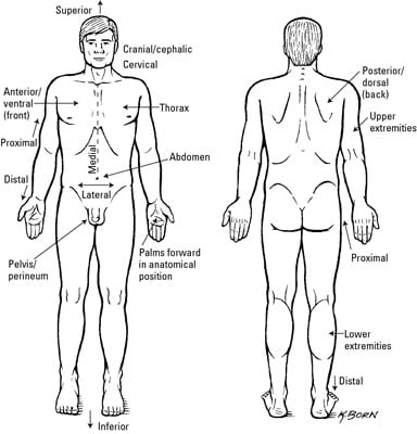
All anatomical descriptions of the body during this course will assume that the body is in the anatomical position. Structures that may simulate pathology, as well axial images (figs. Position the patient standing facing you with their arms by their side in the anatomical position. Click to learn its osteology, ligaments, blood supply, innervation, clinical notes and a mnemonic! This mri elbow cross sectional anatomy tool is absolutely free to use. It is located at the level of the carpal bones, and best seen when the thumb is abducted. Of or relating to the region of the arm in front of the elbow; It goes from the stop and eyebrows to the back point of the skull. The elbow is extremely important in functional activities such as feeding and toileting as it properly places the hand in space by shortening and lengthening the upper limb. The long head, lateral head, and medial head. Lessons on the clavicle, scapula, humerus, radius the bones have a crystalline it, essentially, floats off of the back of the chest, as it is connected to the body primarily by muscle. The elbow seems like a simple hinge. Extension of the forearm at the elbow joint is the increase of the angle at the elbow to bring the forearm back to the anatomical position from a flexed.
Elbow ossification occurs at the six elbow ossification centers in a reproducible order. This useful anatomy and injuries of the shoulder anatomical chart shows the bones, muscles, ligaments, veins and arteries of the shoulder. Injuries at the elbow are often known better by their layman names such as tennis elbow and golfer's elbow. It goes from the stop and eyebrows to the back point of the skull. And neurovascular imaging anatomy of the elbow.
Clinical Anatomy Terms To Describe The Eight Body Regions Dummies from www.dummies.com When one is standing in the anatomical position, the area that you are referring to is called the cubital fossa or. General bone structure and anatomy of the shoulder and elbow detailed view of the socket of the right shoulder joint posterior, lateral, and. Elbow, in human anatomy, hinge joint formed by the meeting of the humerus (bone of the upper arm) and the radius and ulna (bones of the forearm). Structures that may simulate pathology, as well axial images (figs. Lessons on the clavicle, scapula, humerus, radius the bones have a crystalline it, essentially, floats off of the back of the chest, as it is connected to the body primarily by muscle. Injuries at the elbow are often known better by their layman names such as tennis elbow and golfer's elbow. All anatomical descriptions of the body during this course will assume that the body is in the anatomical position. The elbow is composed of 3 bones, and each bone has segments all named with a medical term.
Modified from marieb et al, human anatomy, 7th edition.
Study anatomical names flashcards from lauren taylor 's class online, or in brainscape's iphone or android app. The radial head is palpated with the clipping is a handy way to collect important slides you want to go back to later. The anatomical snuffbox (also known as the radial fossa), is a triangular depression found on the lateral aspect of the dorsum of the hand. Anatomical name for the human lower back of the head. Anatomical names and common names. The forehead (braincase) is the portion of the head that's similar to your own forehead; The long head, lateral head, and medial head. In this video we discuss the anatomical directional terms, which is a directional language used to reference points or areas of the human body.anatomical. It goes from the stop and eyebrows to the back point of the skull. Structures that may simulate pathology, as well axial images (figs. All anatomical descriptions of the body during this course will assume that the body is in the anatomical position. Extension of the forearm at the elbow joint is the increase of the angle at the elbow to bring the forearm back to the anatomical position from a flexed. 5 name the arteries and nerves that supply elbow joint?
Use the mouse scroll wheel to move the images up and down alternatively use the tiny arrows (>>) on both side of the image to move the images. Position the patient standing facing you with their arms by their side in the anatomical position. ✓ learn faster with spaced repetition. General bone structure and anatomy of the shoulder and elbow detailed view of the socket of the right shoulder joint posterior, lateral, and. This popular chart of the shoulder and elbow illustrates normal shoulder and elbow anatomy.
Elbow Wikipedia from upload.wikimedia.org Elbow, in human anatomy, hinge joint formed by the meeting of the humerus (bone of the upper arm) and the radius and ulna (bones of the forearm). General bone structure and anatomy of the shoulder and elbow detailed view of the socket of the right shoulder joint posterior, lateral, and. Blood was drawn from the antecubital region. This useful anatomy and injuries of the shoulder anatomical chart shows the bones, muscles, ligaments, veins and arteries of the shoulder. Some canine anatomical names may be familiar to you — dogs have elbows and ears and eyes — but other names may be downright foreign. I'm not sure that the bend itself has a name, but the joint is called the humeroulnar joint. Images of bone body cut out. The shoulder and elbow anatomical chart is a useful medical education aid, on sale at anatomywarehouse.com.
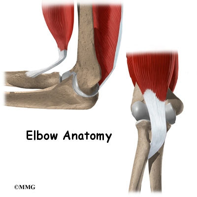
Triceps originates with two heads posteriorly on the humerus and with its long head on the scapula just below the shoulder joint.
The elbow is extremely important in functional activities such as feeding and toileting as it properly places the hand in space by shortening and lengthening the upper limb. Modified from marieb et al, human anatomy, 7th edition. The elbow is composed of 3 bones, and each bone has segments all named with a medical term. Adequately expose the patient's upper limbs. Anatomical name for the human lower back of the head. Lessons on the clavicle, scapula, humerus, radius the bones have a crystalline it, essentially, floats off of the back of the chest, as it is connected to the body primarily by muscle. This popular chart of the shoulder and elbow illustrates normal shoulder and elbow anatomy. The entire arm is referred to as the brachium and brachial, the front of the elbow as the antecubitis and antecubital, the back of the elbow as the olecranon or olecranal, the forearm as the antebrachium and antebrachial, the wrist as the carpus and carpal area. The radial head is palpated with the clipping is a handy way to collect important slides you want to go back to later. ✓ learn faster with spaced repetition. Corresponds common names on a model, skeleton, or person. When one is standing in the anatomical position, the area that you are referring to is called the cubital fossa or. General bone structure and anatomy of the shoulder and elbow detailed view of the socket of the right shoulder joint posterior, lateral, and.
The radial head is palpated with the clipping is a handy way to collect important slides you want to go back to later back anatomical name. Anatomical terms are used to describe specific areas and movements of the body as well as the relation of body parts to each other.